UCLA Researchers Map Mouse Neuron Morphology, Unveil Dendritic Atlas with Aging and Disease Insights
December 4, 2025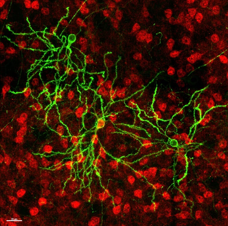
This work creates the first striatum-wide dendritic morphology atlas for these neuron types and demonstrates a systems biology approach linking neuron shape, connectivity, and disease at high resolution.
Looking ahead, the team plans to expand MORF labeling to perturbations and apply MORF plus dendritome mapping to whole-brain tissue clearing and light-sheet imaging, including other neuron types and neurodegenerative diseases.
The analysis covered more than 3,700 D1/D2 MSNs from 12 mice, encompassing both healthy and Huntington's disease model subjects.
UCLA researchers mapped dendritic morphology of two genetically defined mouse striatal neuron types (D1- and D2-MSNs) at unprecedented scale and resolution using MORF labeling, 3D imaging, and computational analysis.
Aging induces dendritic atrophy in both MSN types, while a Huntington's disease model shows modest, differential deficits between D1 and D2 MSNs.
The study finds that D1- and D2-MSNs are broadly similar in morphology, with D1 neurons slightly larger and more complex; regional differences emerge in specific striatal subregions, and MSNs cluster into dendritic modules tied to corticostriatal inputs.
A key methodological advance divides the brain into 7,020 voxels (500 micrometers per side) to generate an eigen-morph per voxel and cluster them into morphometric modules, revealing the spatial organization of dendritic features.
The tools include MORF mice for sparse, bright labeling, thick-tissue imaging with iDISCO clearing, and a pipeline that registers images to the mouse brain atlas (CCFv3) and reconstructs neurons with 31 morphometric features.
Summary based on 1 source
Get a daily email with more Science stories
Source

Medical Xpress • Dec 4, 2025
Comprehensive map reveals neuronal dendrites in the mouse brain in greater detail