Revolutionary LICONN Method Enhances Brain Tissue Imaging Using Standard Microscopes
May 7, 2025
The integration of computer science and deep learning techniques from Google Research has been crucial in processing the vast data generated, automating the identification of neurons and their connections.
One of the key advantages of this method is its ability to identify molecules, cells, and their connections without relying on costly electron microscopes.
LICONN, a groundbreaking microscopy method developed by scientists at the Institute of Science and Technology Austria (ISTA) in collaboration with Google Research, enables detailed reconstruction of brain tissue and synaptic connections.
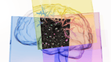
This innovative technique employs hydrogel expansion to enhance imaging resolution, allowing for high-fidelity visualization of neuronal structures such as axons and dendritic spines.
Traditional light microscopes typically achieve a resolution of only 250-300 nanometers, which is inadequate for observing the densely packed structures found in brain tissue.
The study successfully applied immunolabelling techniques to synaptic proteins like bassoon and PSD95, shedding light on the molecular architecture of synapses within the reconstructed tissue.
LICONN utilizes standard light microscopes, making it an accessible and reproducible solution for researchers worldwide, eliminating the need for expensive equipment.
The method has been validated against ground truth datasets, demonstrating high accuracy in tracing axons and dendrites, with a significant percentage of spines correctly linked to their parent dendrites.
This multidisciplinary approach brings researchers closer to unraveling the intricate workings of the mammalian brain and its functions in both health and disease.
Initial applications of LICONN have focused on mapping mouse brain tissue, with future plans to extend its use to human brains, enhancing our understanding of brain connectivity.
LICONN is poised to facilitate routine connectomic studies in non-specialized laboratories and has potential applications for high-resolution tissue analysis in other organs.
Published in the journal Nature, this method represents a significant advancement in visualizing the brain's complex networks without the necessity of expensive electron microscopy.
Summary based on 8 sources
Get a daily email with more AI stories
Sources
Google • May 7, 2025
Google Research and ISTA are using light microscopes to "map" the brain.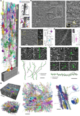
Nature • May 7, 2025
Light-microscopy-based connectomic reconstruction of mammalian brain tissue
ScienceDaily • May 7, 2025
Piecing together the brain puzzle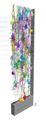
Phys.org • May 7, 2025
Microscopy method can reconstruct mammalian brain tissue in synaptic detail