Breakthrough 3D Imaging Reveals How Human Embryos Implant and Connect in Real-Time
August 15, 2025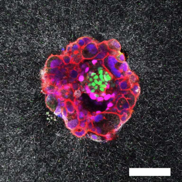
Researchers have captured the first real-time, three-dimensional images and videos of human embryos implanting into a collagen-based tissue model that mimics the uterus, providing unprecedented insights into the mechanics of implantation.
Unlike mouse embryos, human embryos are larger, more invasive, and actively penetrate the uterine lining by releasing enzymes, exerting force, and connecting with maternal blood vessels.
The study shows that embryo forces and tissue interactions, including enzyme release and traction, are critical for successful implantation, which influences fertility and miscarriage rates.
This research extends the observation window beyond the initial five days post-fertilization, allowing scientists to monitor earlier and later stages of embryo development, and highlights the importance of mechanical forces and matrix displacement.
Experts emphasize that these findings lay the groundwork for future studies on implantation mechanisms and could lead to new treatments for implantation failure.
While the current collagen matrix isn't intended for IVF, it could be useful for pharmaceutical testing and understanding embryo behavior, potentially aiding in embryo selection.
The developed platform can be used in both 2D and 3D formats to test environmental conditions or compounds, such as a protein supplement aimed at improving implantation success rates.
Future research will focus on standardizing experimental materials and encouraging embryo donation for research, made possible by donor generosity.
This breakthrough provides dynamic visualization of early development stages that were previously only observable through snapshots or ultrasound, revealing how embryos invade and connect within a collagen matrix.
Understanding the mechanical forces and contractions involved in implantation could lead to improved fertility treatments, as failure to implant accounts for about 60% of miscarriages.
External tension and contractions may guide embryos toward blood vessels and nutrients, with some evidence suggesting an optimal frequency of uterine contractions for successful implantation.
Human embryos donated by a hospital were observed over 16-24 hours, with images captured every 20 minutes to document the implantation process.
This research provides detailed visual data on how human embryos invade a collagen-based matrix, revealing the dynamic mechanics of early implantation stages.
Summary based on 7 sources
Get a daily email with more World News stories
Sources

The Guardian • Aug 15, 2025
Scientists capture first footage of human embryo implanting in a uterus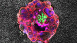
Nature • Aug 15, 2025
Watch a human embryo implant itself — with brute force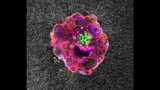
Gizmodo • Aug 15, 2025
In a First, Scientists Capture Human Embryo Implantation in Real Time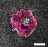
Scientific American • Aug 15, 2025
Human Embryo Implantation Revealed in First-Ever 3D Images