Revolutionary Cryo-Optical Microscopy Captures Cellular Activities with Millisecond Precision
August 23, 2025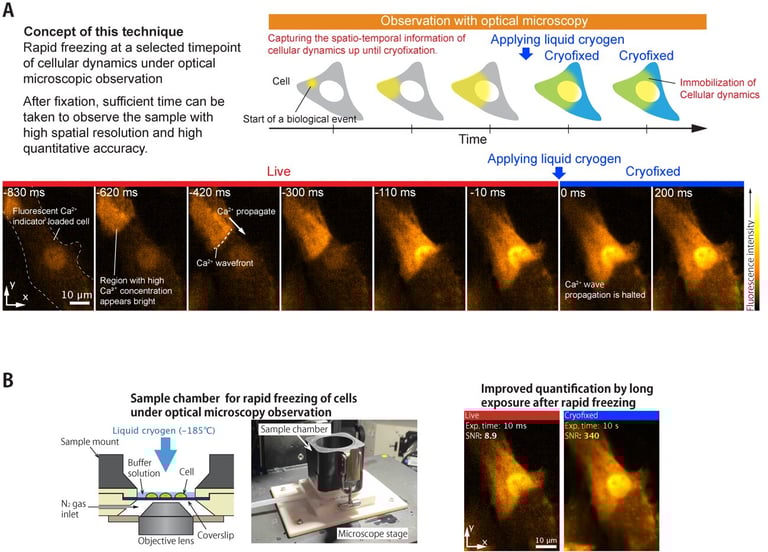
The development includes a custom on-stage freezing chamber that minimizes ice crystal formation and maintains low drift velocities, ensuring high-resolution imaging under cryogenic conditions.
This cryo-approach enhances optical stability and reduces photobleaching, significantly improving the sensitivity and accuracy of quantitative fluorescence imaging at nanomolar precision.
Published in Light: Science & Applications, this research highlights its potential to transform our understanding of dynamic biological processes, offering new insights for life sciences and medical research.
Researchers from The University of Osaka have developed a groundbreaking cryo-optical microscopy technique that allows rapid, in situ freezing of living cellular samples, capturing high-resolution snapshots of cellular activity at specific moments with millisecond precision.
This method effectively halts rapid cellular activities like calcium ion wave propagation in heart muscle cells, capturing intricate details with super-resolution techniques at different stages with 10 ms precision.
An electrically triggered cryogen injection system with 10 ms timing accuracy was used to freeze transient biological events, such as calcium release induced by UV light, at specific moments.
By freezing cells labeled with fluorescent probes, the technique enables longer exposure times—up to 1,000 times longer than live imaging—resulting in more accurate quantitative measurements without motion blur.
The technique supports multimodal imaging, combining methods like Raman microscopy and super-resolution fluorescence microscopy, in a single frozen sample for comprehensive, synchronized analysis.
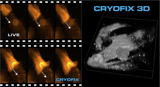
It also enables detailed 3D super-resolution imaging of subcellular structures at the exact moment of biological events, facilitating the study of dynamic cellular processes previously difficult to capture.
This innovative method preserves cellular structures and dynamics under cryogenic conditions, opening new avenues for studying fast, transient cellular phenomena in cell biology, biophysics, and medicine.
Senior author Katsumasa Fujita emphasized that this technique shifts the paradigm from tracking fast cellular movements to arresting cellular processes for detailed study, opening new possibilities for scientific discovery.
The approach shifts the focus from real-time tracking of rapid cellular processes to arresting these processes at precise moments, allowing detailed analysis of transient events such as calcium waves.
Summary based on 4 sources
Get a daily email with more Science stories
Sources
Nature • Aug 23, 2025
Time-deterministic cryo-optical microscopy
Phys.org • Aug 23, 2025
Freeze-framing the cellular world to capture a fleeting moment of activity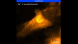
Cosmos • Aug 23, 2025
Push pause to capture high-resolution snapshots of cells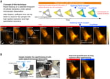
Asia Research News • Aug 23, 2025
Freeze-framing the cellular world to capture a fleeting moment of cellular activity