Breakthrough 3D Imaging of Cardiac Ryanodine Receptors Unveiled with MINFLUX Microscopy
December 21, 2025
The method revealed the subunit architecture and 3D orientation of RyR in living cardiomyocytes, highlighting unprecedented detail in situ.
These findings may inform future therapies targeting cardiac function by deepening molecular-level knowledge of RyR structure and organization.
Researchers Clowsley, Meletiou, Janicek, and collaborators conducted the study to uncover the precise orientation and structural organization of RyR.
The study provides detailed visualization of RyR architecture, offering new insights into its role in cardiac excitation-contraction coupling.
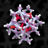
MINFLUX microscopy mapped the three-dimensional structure and subunit arrangement of the cardiac ryanodine receptor (RyR) within living cells, achieving nanometer-scale resolution in this context.
The work reveals distinct RyR subunit clustering and conformational heterogeneity that correlate with functional states within the cell’s 3D environment.
Findings enable in situ differentiation of RyR-subunit interactions with regulatory proteins and suggest how structural variations may influence calcium signaling in health and disease, including arrhythmias and heart failure.
MINFLUX’s robustness in cellular systems is demonstrated, addressing challenges like fluorophore density, background noise, and autofluorescence, with computational algorithms refining localization data.
RyR subunits were labeled with fluorescent probes and localized sequentially under cryogenic conditions to stabilize structures and boost accuracy, leveraging MINFLUX’s photon-efficient detection.
Three-dimensional reconstructions showed tilts and rotations of RyR subunits relative to the sarcoplasmic reticulum membrane, indicating dynamic conformational plasticity linked to gating mechanisms.
Future directions include longitudinal imaging of RyR conformational changes, integrating with other modalities, and extending the approach to in vivo models and human cardiac tissue to validate clinical relevance.
The cardiac ryanodine receptor governs calcium release during contraction, and achieving live-cell imaging at molecular resolution advances understanding of RyR function.
Summary based on 2 sources