New Study Maps Right Ventricle's Cellular Changes, Reveals Insights into Heart Failure Progression
October 9, 2026

Right ventricular failure unfolds in two stages: an initial phase of broad transcriptional remodeling and loss of protective programs across multiple cell lineages before functional decompensation, followed by a second phase with cell-type-specific programs, notably BMP/TGFβ signaling in cardiomyocytes and EndoMT/angiogenic destabilization in endothelial cells.
A two-phase model of RV failure progression is proposed: Phase 1 NF to pRV involves widespread erosion of protective programs across diverse cell lineages, while Phase 2 pRV to RVF features fewer global changes and more targeted, cell-type-specific remodeling.
Immune profiling identifies an inflammatory macrophage population with immediate-early gene expression and resident macrophage markers, confirmed across single-nucleus and spatial transcriptomics, indicating a tissue-specific immune landscape distinct from left-ventricle atlases.
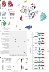
Single-nucleus RNA-seq resolves 34 cell subtypes across 12 lineages, with fibroblasts showing the strongest transcriptional response and substantial differentially expressed genes in pRV and RVF, while cardiomyocytes also exhibit notable changes including mitochondrial gene downregulation and reactivation of fetal programs.
Cross-species analysis shows mouse a model of pulmonary artery banding replicates metabolic and structural features but misses the human-specific antigen presentation program; pediatric hypoplastic left heart syndrome hearts resist mitochondrial collapse and preserve respiratory capacity despite high afterload.
Across etiologies, RVF shows shared mitochondrial decline and upregulated interferon signaling, while complement cascade responses appear etiology-specific, highlighting both common and distinct pathways in right-ventricle pathology.
Translational implications include proposing anti-fibrotic strategies targeting TGFBR3, immunomodulatory approaches, and direct mitochondrial support as potential RV-specific therapies, with fibrosis-related transcripts serving as remodeling biomarkers.
The atlas and analysis pipeline are publicly deposited, enabling broad use to understand pediatric and adult right-ventricle failure and to guide future therapeutic development.
WGCNA reveals divergent modules: oxidative phosphorylation declines, an adaptive immune module rises, and a resident macrophage module drops with disease progression, with concordant signals across platforms.
Cell–cell communication analysis shows stage-specific remodeling, with baseline adhesion pathways maintained but RVF featuring a gain of ECM signaling (COLLAGEN, LAMININ, THBS), indicating enhanced fibroblast-to-stroma interactions during remodeling.
The study creates the most detailed molecular atlas of the human right ventricle by integrating bulk RNA-seq, snRNA-seq, and spatial transcriptomics across healthy to failing tissue, mapping 34 cell subtypes in 12 lineages.
An integrated multimodal atlas of the RV tracks progression from normal function to pressure-loaded preserved function (pRV) and failing RV (RVF), incorporating bulk RNA-seq, snRNA-seq, Xenium spatial transcriptomics, mitochondria respirometry, flow cytometry and histology.
Summary based on 2 sources
Get a daily email with more Science stories
Sources

BIOENGINEER.ORG • Oct 9, 2026
Landmark heart atlas reveals two-phase collapse of the failing right