Groundbreaking Atlas Maps Human Meninges Development from Embryo to Fetus, Highlights Trisomy 21 Effects
September 28, 2026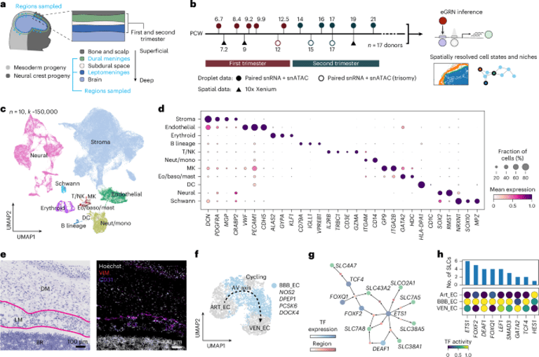

A comprehensive single-cell and spatial atlas of the developing human meninges tracks embryonic week 5 to fetal weeks 9–13, integrating scRNA-seq, spatial transcriptomics, and analyses of published meningioma data to map cellular programs.
The atlas defines a transcriptional and epigenetic framework for meningeal development, highlighting fibroblast lineages, blood–brain barrier endothelium, myeloid and B cell lineages, and notes how trisomy 21 can influence these programs.
Layer identity is delineated: pia marked by LAMC3, arachnoid by SLC22A6, dura by COL8A1, with inner dura distinguished by SLC47A1 and outer dura by MSX2, plus a putative common skull/dura progenitor around the primary meninx.
Spatial fibroblast mapping reveals seven fine-grained states (e.g., Pia_FIB, Arach_FIB, Dura_FIB, Cycling_FIB) and a small pre-osteoblast population, suggesting a developmental progression from Pia_FIB and Cycling_FIB to Arach_FIB and Dura_FIB.
Meningeal myeloid niches include progenitors, meningeal macrophages, and LYVE1-expressing BAMs, with yolk sac–like origins and spatial patterns placing BAM near vasculature and MG deeper in the brain, plus TGFB1/IGF1 signaling indicating potential angiogenic roles for BAM_FOLR2low.
Vasculature and choroid plexus evolve from a primary vascular plexus to arterial/venous capillaries and eventual arterioles, with early venous lymphatic markers PROX1 suggesting subarachnoid lymphangiogenesis.
Blood–brain barrier endothelial development is characterized by arterial, venous, and cycling states, with ETS1-regulated transport networks and key transporters like SLC7A5, SLC7A8, and SLC43A2 highlighted.
Inner dura exhibits tight-junction–related gene expression, and eight spatial clusters map the meninges in the fetal head, including inner/outer dura, arachnoid, pia, and skull/skin fibroblast populations.
The atlas identifies major cell superclasses in the meninges—immune, vascular, and fibroblast—with dominant FOXC1+ fibroblasts and extensive endothelial, perivascular, immune, erythropoietic, neural, neural crest, and epithelial cell types characterized.
In trisomy 21 meninges, a gene-dosage effect emerges with widespread differential expression; stromal and erythroid cells are depleted, Pia_FIB is disproportionately affected, and JAK–STAT, KRAS, and MTORC1 signaling are upregulated in immune compartments while stromal chemokine signaling is downregulated.
Choroid plexus development shows epithelial differentiation and neural–epithelial progenitors, with human-specific fibroblast features suggesting immune surveillance at the blood–CSF border.
Immune development is unexpectedly diverse in the fetal meninges, with 19 immune cell types identified; B-lineage cells appear early and mature from pre- to immature B cells into later stages by mid-gestation, indicating prenatal establishment of meningeal B cell compartments.
Summary based on 2 sources