New Atlas Maps Postnatal Heart Development, Reveals Cardiomyocyte Maturation Dynamics
September 8, 2026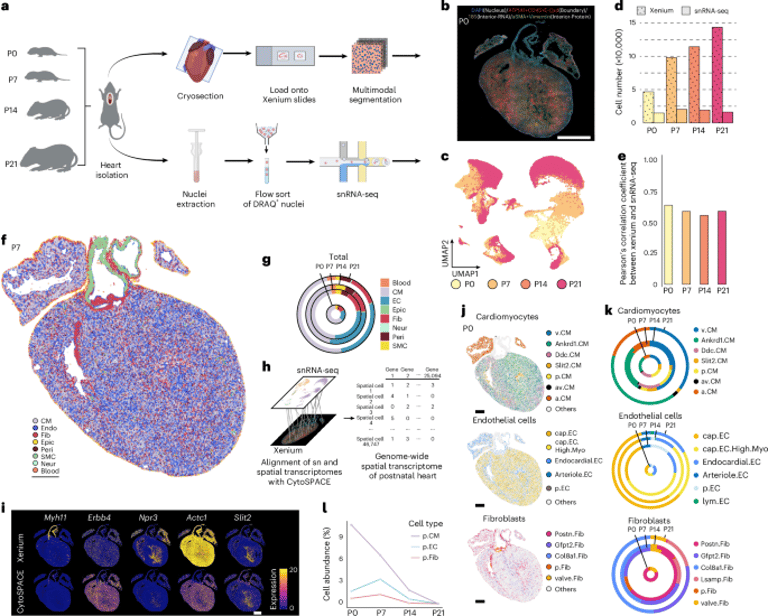

A comprehensive study combines single-nucleus transcriptomics with spatial transcriptomics to define cellular niches and regulatory networks driving postnatal cardiomyocyte maturation, mapping how gene programs unfold across time and space.
Using a spatially guided in vivo single-cell functional genomics approach, the team builds a high-resolution spatiotemporal atlas of the heart at P0, P7, P14 and P21, delineating postnatal heart development.
Readers should note that the content is a preview or subscription-based article, with options to read or purchase the full article, and references a related full-length paper by Wang et al.
A notable cardiomyocyte subpopulation, Ankrd1.CMs, is enriched in the left ventricle and peri-arteriolar regions, showing stress-response gene upregulation that suggests mechanosensitive maturation states during postnatal growth.
SigNET is introduced as a pipeline linking signaling pathways to transcriptional networks, capturing downstream transcription factors like Elk3 as mediators of Kitl−Kit–driven endothelial sprouting during capillary plexus expansion and supporting Kitl−Kit’s role in postnatal angiogenesis.
The findings are contextualized within existing literature on cardiomyocyte maturation, noting related prior work and reviews cited in the references.
Network analysis of 197 ligand–receptor pairs reveals spatially constrained intercellular signaling that shifts from Postn.Fibs dominance early (P0–P7) to cap.ECs dominance by P14, coordinating maturation processes.
The atlas identifies eight major cell types and resolves 25 transcriptionally and spatially distinct cellular states with specific regional localization patterns across cardiomyocytes, endothelial cells and fibroblasts.
Maturation signals in cardiomyocytes are quantified via AUC analyses, using a Xenium probe set to monitor maturation-associated genes and transcription factors within spatial contexts, highlighting region-specific maturation dynamics.
Spatially resolved regulon analysis via SCENIC shows stage-specific transcription factor activity in cardiomyocytes, with non-myocytes showing more stable regulon profiles; TFs such as Fosl2 and Foxc1 emerge as potential regulators in epicardial and chamber development.
A public web atlas (postnatalheartatlas.org) enables interactive exploration of gene expression, regulon activity and cell communication dynamics at single-cell resolution, broadening access to the dataset.
The study defines 12 cellular niches aligned with anatomical structures, with niches showing dynamic composition and proliferative states as activity shifts from early postnatal stages toward maturation.
Summary based on 2 sources
Get a daily email with more Science stories
Sources
Nature • Sep 7, 2026
Spatially guided in vivo single-cell functional genomics of postnatal heart